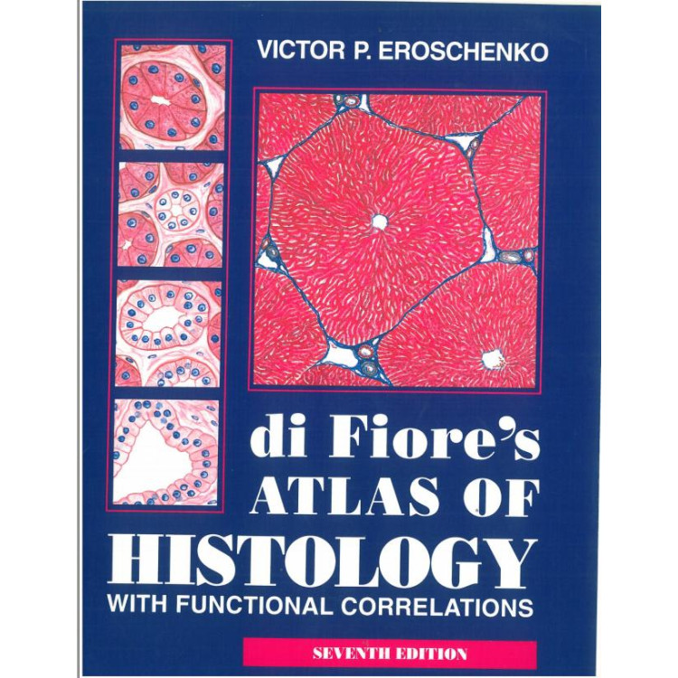
di Fiores Atlas of Histology with functional correlations Eroschenko
Сумата се прибавя директно в кошницата
12.24 €
Етикети: di Fiores Atlas of Histology with functional correlations Eroschenko
PLATE 1 FIG. 1. FIG. 2. FIG. 3. Simple Squamous Epithelium: Dissociated Squamous Epithelial Cells Simple Squamous Epithelium: Surface View of Peritoneal Mesothelium Simple Squamous Epithelium: Peritoneal Mesothelium (Traverse Section)
PLATE 2
FIG. J. FIG. 2. Simple Columnar Epithelium Simple Columnar Epithelium: Cells with Striated Borders and Goblet Cells
PLATE 3 FIG. 1. FIG. 2. Stratified Squamous Epithelium (Transverse Section) Stratified Squamous Epithelium (Tangential Section)
PLATE 4 FIG. 1. FIG. 2. Pseudostratified Columnar Ciliated Epithelium Transitional Epithelium
PLATE 5 Simple Branched Tubular Gland (Diagram)
PLATE 6 Compound Tubuloacinar Gland (Diagram)
PLATE 7 Compound Acinar Gland (Diagram)
THE CONNECTIVE TISSUE
PLATE 8 FIG. 1. FIG. 2. Loose Irregular Connective Tissue (Spread) Cells of Loose Connective Tissue in Sections
PLATE 9 FIG. 1. FIG. 2. Loose Irregular Connective Tissue Dense Irregular Connective Tissue
PLATE 1 0 FIG. 1. FIG. 2. Dense Regular Connective Tissue: Tendon (Longitudinal Section) Dense Regular Connective Tissue: Tendon [Transverse Section)
PLATE 1 1 FIG. J. FIG. 2. FIG. 3. Dense Irregular and Loose Connective Tissue (Elastin Stain) Adipose Tissue Embryonic Connective Tissue
CONTENTS
IFIG. 1. FIG. 1 A. FIG. 2. FIG. 3. Fetal Cartilage: Developing Hyaline Cartilage Fetal Cartilage (Sectional ViewJ Mature Hyaline Cartilage Newly Formed Hyaline Cartilage of the Trachea
PLATE 1 3 FIG. 1. FIG. 2. Fibrous Cartilage: Intervertebral Disk Elastic Cartilage: Epiglottis
THE BONE
PLATE 1 4 FIG. 1. FIG. 2. Compact Bone, Dried: Diaphysis of the Tibia (Transverse Section) Compact Bone, Dried: Diaphysis of the Tibia (Longitudinal Section)
PLATE 1 5 FIG. 1. FIG. 2. Cancellous Bone: Adult Sternum (Transverse Section, Decalcified) Intramembranous Ossification: Mandible of Five-Month Fetus (Transverse Section, Decalcified)
PLATE 1 6 Endochondral Ossification: Developing Metacarpal Bone (Panoramic View, Longitudinal Section)
PLATE 1 7 Endochondral Ossification (Sectional View)
PLATE 1 8 Formation of Bone: Development of Osteons (Haversian Systems) (Transverse Section, Decalcified),
PLATE 1 9 Formation of Bone: Secondary (Epiphyseal) Ossification Centers (Longitudinal Section, Decalcified),
THE BLOOD
PLATE 20 Peripheral Blood Smear
PLATE 2 1 FIG. 1. FIG. 2. Blood Cells: Supravital Stains Blood Smears: Pappenheim's and Celani's Stains
PLATE 22 FIG. 1. FIG. 2. Hemopoietic Bone Marrow Bone Marrow of a Rabbit, India Ink Preparation
PLATE 23 Bone Marrow: Smear