Radiology, Marco Rau - теми
Сумата се прибавя директно в кошницата
4.08 €
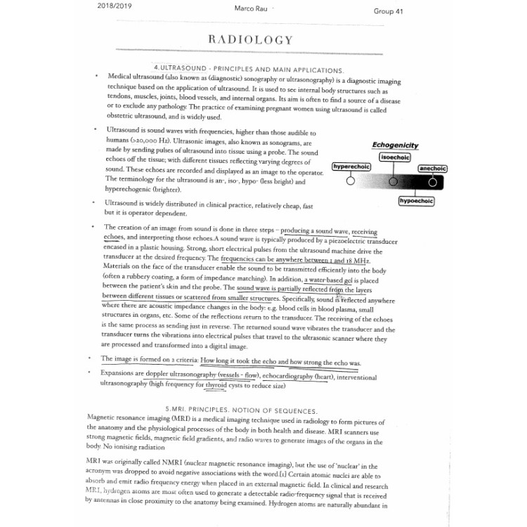
RADIOLOGY
4.ULTRASOUND - PRINCIPLES AND MAIN APPLICATIONS.
* Medical ultrasound (also known as (diagnostic) sonography or ultrasonography) is a diagnostic imaging technique based on the application of ultrasound. It is used to see internal body structures such as tendons, muscles, joints, blood vessels, and internal organs. Its aim is often to find a source of a disease or to exclude any pathology The practice of examining pregnant women using ultrasound is called obstetric ultrasound, and is widely used.
* Ultrasound is sound waves with frequencies, higher than those audible to humans (>20,000 Hz). Ultrasonic images, also known as sonograms, are made by sending pulses of ultrasound into tissue using a probe. The sound echoes off the tissue; with different tissues reflecting varying degrees of sound. These echoes are recorded and displayed as an image to the operator.
The terminology for the ultrasound is an-, iso-, hypo- (less bright) and hyperechogenic (brighter).
* Ultrasound is widely distributed in clinical practice, relatively cheap, fast but it is operator dependent.
* The creation of an image from sound is done in three steps - producing a sound wave, receiving echoes, and interpreting those echoes.A sound wave is typically produced by a piezoelectric transducer encased in a plastic housing. Strong, short electrical pulses from the ultrasound machine drive the transducer at the desired frequency. The frequencies can be anywhere between 1 and r8 MHz.
Materials on the face of the transducer enable the sound to be transmitted efficiently into the body (often a rubbery coating, a form of impedance matching). In addition, a waterbased gel is placed between the patient's skin and the probe. The sound wave is partially reflected from :he layers between different tissues or scattered from smaller structures. Specifically, sound is reflected anywhere where there are acoustic impedance changes in the body: e.g. blood cells in blood plasma, small structures in organs, etc. Some of the reflections return to the transducer. The receiving of the echoes is the same process as sending just in reverse. The returned sound wave vibrates the transducer and the transducer turns the vibrations into electrical pulses that travel to the ultrasonic scanner where they are processed and transformed into a digital image.